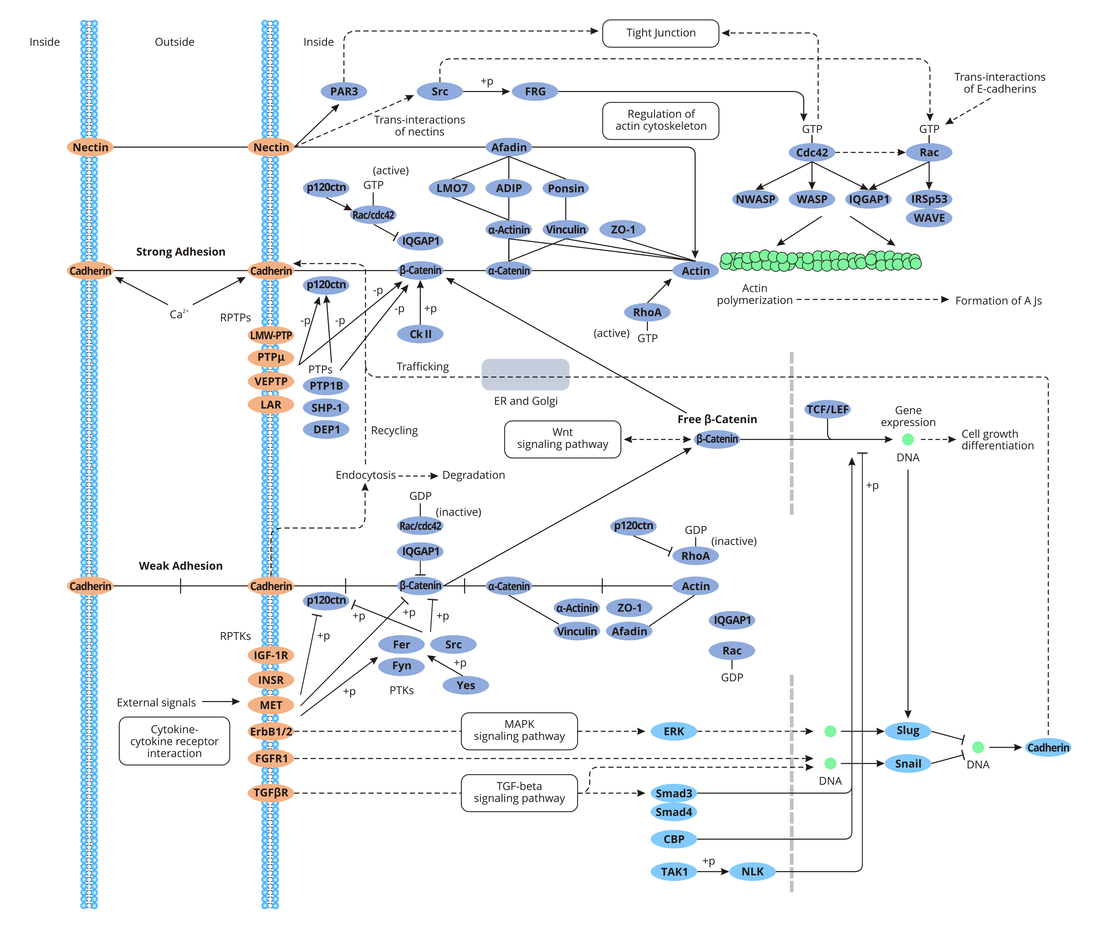
Adherens Junction Diagram . They hold cardiac muscle cells tightly together as the heart expands and contracts and hold epithelial cells together. tight junctions (blue dots) between cells are connected areas of the plasma membrane that stitch cells together. The adherens junction lies below the tight junction (occluding junction). adherens junctions provide strong mechanical attachments between adjacent cells. adherens junctions are positioned immediately below tight junctions and characterized by two apposing membranes, which are separated by ∼20 n. Adherens junctions (red dots) join the actin filaments of neighboring cells. adherens junctions are built primarily from cadherins, whose extracellular segments bind to each other and whose intracellular segments bind to. epithelial cells are held together by strong anchoring (zonula adherens) junctions.
from www.cusabio.com
They hold cardiac muscle cells tightly together as the heart expands and contracts and hold epithelial cells together. Adherens junctions (red dots) join the actin filaments of neighboring cells. adherens junctions are positioned immediately below tight junctions and characterized by two apposing membranes, which are separated by ∼20 n. epithelial cells are held together by strong anchoring (zonula adherens) junctions. adherens junctions are built primarily from cadherins, whose extracellular segments bind to each other and whose intracellular segments bind to. tight junctions (blue dots) between cells are connected areas of the plasma membrane that stitch cells together. adherens junctions provide strong mechanical attachments between adjacent cells. The adherens junction lies below the tight junction (occluding junction).
Adherens Junction CUSABIO
Adherens Junction Diagram adherens junctions provide strong mechanical attachments between adjacent cells. epithelial cells are held together by strong anchoring (zonula adherens) junctions. adherens junctions are built primarily from cadherins, whose extracellular segments bind to each other and whose intracellular segments bind to. Adherens junctions (red dots) join the actin filaments of neighboring cells. adherens junctions are positioned immediately below tight junctions and characterized by two apposing membranes, which are separated by ∼20 n. adherens junctions provide strong mechanical attachments between adjacent cells. tight junctions (blue dots) between cells are connected areas of the plasma membrane that stitch cells together. The adherens junction lies below the tight junction (occluding junction). They hold cardiac muscle cells tightly together as the heart expands and contracts and hold epithelial cells together.
From www.researchgate.net
Endothelial cell tight junctions and adherens junction proteins. The Adherens Junction Diagram adherens junctions are built primarily from cadherins, whose extracellular segments bind to each other and whose intracellular segments bind to. They hold cardiac muscle cells tightly together as the heart expands and contracts and hold epithelial cells together. adherens junctions are positioned immediately below tight junctions and characterized by two apposing membranes, which are separated by ∼20 n.. Adherens Junction Diagram.
From www.researchgate.net
Adherens junction components and their structural (A), signal Adherens Junction Diagram adherens junctions are positioned immediately below tight junctions and characterized by two apposing membranes, which are separated by ∼20 n. tight junctions (blue dots) between cells are connected areas of the plasma membrane that stitch cells together. Adherens junctions (red dots) join the actin filaments of neighboring cells. They hold cardiac muscle cells tightly together as the heart. Adherens Junction Diagram.
From www.researchgate.net
Summary of the interactions of nuclear adherens junction proteins Adherens Junction Diagram adherens junctions provide strong mechanical attachments between adjacent cells. tight junctions (blue dots) between cells are connected areas of the plasma membrane that stitch cells together. Adherens junctions (red dots) join the actin filaments of neighboring cells. The adherens junction lies below the tight junction (occluding junction). They hold cardiac muscle cells tightly together as the heart expands. Adherens Junction Diagram.
From www.researchgate.net
Adherens junction components are enriched at rosette centers ac Adherens Junction Diagram They hold cardiac muscle cells tightly together as the heart expands and contracts and hold epithelial cells together. adherens junctions are built primarily from cadherins, whose extracellular segments bind to each other and whose intracellular segments bind to. The adherens junction lies below the tight junction (occluding junction). Adherens junctions (red dots) join the actin filaments of neighboring cells.. Adherens Junction Diagram.
From www.researchgate.net
Adherens junction assembly (cadherins) pathway using gene set Adherens Junction Diagram They hold cardiac muscle cells tightly together as the heart expands and contracts and hold epithelial cells together. adherens junctions provide strong mechanical attachments between adjacent cells. adherens junctions are positioned immediately below tight junctions and characterized by two apposing membranes, which are separated by ∼20 n. tight junctions (blue dots) between cells are connected areas of. Adherens Junction Diagram.
From www.researchgate.net
Structure of anchoring junctions. Adherens junctions are attached to Adherens Junction Diagram adherens junctions provide strong mechanical attachments between adjacent cells. adherens junctions are built primarily from cadherins, whose extracellular segments bind to each other and whose intracellular segments bind to. They hold cardiac muscle cells tightly together as the heart expands and contracts and hold epithelial cells together. tight junctions (blue dots) between cells are connected areas of. Adherens Junction Diagram.
From wirelibrotheomanias.z13.web.core.windows.net
Tight Junctions Diagram Adherens Junction Diagram adherens junctions provide strong mechanical attachments between adjacent cells. adherens junctions are built primarily from cadherins, whose extracellular segments bind to each other and whose intracellular segments bind to. Adherens junctions (red dots) join the actin filaments of neighboring cells. They hold cardiac muscle cells tightly together as the heart expands and contracts and hold epithelial cells together.. Adherens Junction Diagram.
From www.researchgate.net
6 Scatter plot of normalised expression of adherens junction pathway Adherens Junction Diagram epithelial cells are held together by strong anchoring (zonula adherens) junctions. tight junctions (blue dots) between cells are connected areas of the plasma membrane that stitch cells together. adherens junctions are built primarily from cadherins, whose extracellular segments bind to each other and whose intracellular segments bind to. Adherens junctions (red dots) join the actin filaments of. Adherens Junction Diagram.
From exohekjkc.blob.core.windows.net
Communicating Junctions Definition at Anita Richards blog Adherens Junction Diagram adherens junctions provide strong mechanical attachments between adjacent cells. They hold cardiac muscle cells tightly together as the heart expands and contracts and hold epithelial cells together. The adherens junction lies below the tight junction (occluding junction). Adherens junctions (red dots) join the actin filaments of neighboring cells. tight junctions (blue dots) between cells are connected areas of. Adherens Junction Diagram.
From quizlet.com
Adherens Junctions Diagram Quizlet Adherens Junction Diagram They hold cardiac muscle cells tightly together as the heart expands and contracts and hold epithelial cells together. adherens junctions provide strong mechanical attachments between adjacent cells. tight junctions (blue dots) between cells are connected areas of the plasma membrane that stitch cells together. The adherens junction lies below the tight junction (occluding junction). Adherens junctions (red dots). Adherens Junction Diagram.
From www.researchgate.net
The junctional complexes of the intestinal barrier. Tight junctions are Adherens Junction Diagram tight junctions (blue dots) between cells are connected areas of the plasma membrane that stitch cells together. adherens junctions are positioned immediately below tight junctions and characterized by two apposing membranes, which are separated by ∼20 n. epithelial cells are held together by strong anchoring (zonula adherens) junctions. adherens junctions are built primarily from cadherins, whose. Adherens Junction Diagram.
From dxopflzpo.blob.core.windows.net
Adherens Junction Proteins at Lamar Moody blog Adherens Junction Diagram The adherens junction lies below the tight junction (occluding junction). epithelial cells are held together by strong anchoring (zonula adherens) junctions. They hold cardiac muscle cells tightly together as the heart expands and contracts and hold epithelial cells together. tight junctions (blue dots) between cells are connected areas of the plasma membrane that stitch cells together. adherens. Adherens Junction Diagram.
From www.geeksforgeeks.org
Gap Junction Definition, Structure, Functions and FAQs Adherens Junction Diagram They hold cardiac muscle cells tightly together as the heart expands and contracts and hold epithelial cells together. adherens junctions are positioned immediately below tight junctions and characterized by two apposing membranes, which are separated by ∼20 n. tight junctions (blue dots) between cells are connected areas of the plasma membrane that stitch cells together. The adherens junction. Adherens Junction Diagram.
From www.researchgate.net
Major molecules of the tight and adherens junctions are shown. Tight Adherens Junction Diagram Adherens junctions (red dots) join the actin filaments of neighboring cells. The adherens junction lies below the tight junction (occluding junction). adherens junctions are positioned immediately below tight junctions and characterized by two apposing membranes, which are separated by ∼20 n. adherens junctions provide strong mechanical attachments between adjacent cells. adherens junctions are built primarily from cadherins,. Adherens Junction Diagram.
From www.researchgate.net
Major molecules of the tight and adherens junctions are shown. Tight Adherens Junction Diagram The adherens junction lies below the tight junction (occluding junction). epithelial cells are held together by strong anchoring (zonula adherens) junctions. adherens junctions provide strong mechanical attachments between adjacent cells. adherens junctions are positioned immediately below tight junctions and characterized by two apposing membranes, which are separated by ∼20 n. tight junctions (blue dots) between cells. Adherens Junction Diagram.
From med.libretexts.org
4.2B Adherens Junctions Medicine LibreTexts Adherens Junction Diagram adherens junctions are built primarily from cadherins, whose extracellular segments bind to each other and whose intracellular segments bind to. tight junctions (blue dots) between cells are connected areas of the plasma membrane that stitch cells together. adherens junctions provide strong mechanical attachments between adjacent cells. They hold cardiac muscle cells tightly together as the heart expands. Adherens Junction Diagram.
From www.geeksforgeeks.org
Adherens Junction Structure, Functions, Examples, and FAQs Adherens Junction Diagram tight junctions (blue dots) between cells are connected areas of the plasma membrane that stitch cells together. Adherens junctions (red dots) join the actin filaments of neighboring cells. They hold cardiac muscle cells tightly together as the heart expands and contracts and hold epithelial cells together. epithelial cells are held together by strong anchoring (zonula adherens) junctions. . Adherens Junction Diagram.
From www.alamy.com
Cell junctions tight junction (or occluding), adherens junction Stock Adherens Junction Diagram epithelial cells are held together by strong anchoring (zonula adherens) junctions. adherens junctions are positioned immediately below tight junctions and characterized by two apposing membranes, which are separated by ∼20 n. tight junctions (blue dots) between cells are connected areas of the plasma membrane that stitch cells together. adherens junctions are built primarily from cadherins, whose. Adherens Junction Diagram.
